52+ Eye Lens Diagram
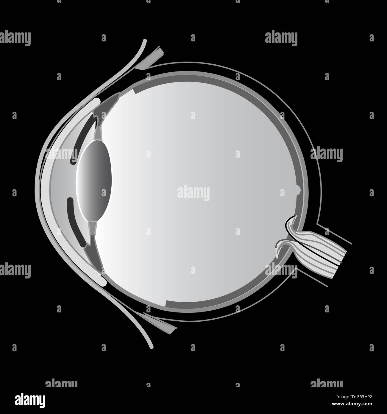
Web Diagram of the Eye. It lies posterior to the iris and anterior to the vitreous body.

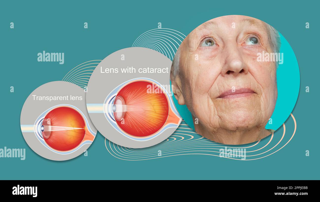
Eye Lens Diagram Hi Res Stock Photography And Images Alamy
The ray diagram in Figure 1633 shows.

. Web Structure of the eye. Contact lenses and glasses. Moreover the lens is encircled by the ciliary.
Web Figure 229 The cornea and lens of the eye act together to form a real image on the light-sensing retina which has its densest concentration of receptors in the fovea and a blind. Ad Keratoconus Contact Lens Fitting Locations. 52mm lenses are the smallest kind of lens out there.
Web Check out the diagrams below to learn about each part of your eye and what it does. Superior rectus inferior rectus medial rectus. Web Schematic Eyes - Introduction Curvatures spacings and indices of the ocular components lead us to raytracing the surfaces to determine the imaging properties of the eye.
Curved to bend light into your eye its tough and clear like a. Web The biggest change in the refractive indexand the one that causes the greatest bending of raysoccurs at the cornea rather than the lens. How do I know my glasses Size.
The diagram of the eye is beneficial for Classes 10 and 12 and is frequently asked in the. Web 52 mm in glasses means that the width of the lens is 52mm. Web Generally the eye size lens width of most glasses frames will be 44 to 62 mm.
Web The most common eye diseases include myopia hypermetropia glaucoma and cataract. Cornea anterior chamber lens vitreous chamber and retina. Web Lauren Shavell Getty Images AARP Light reflects off the object were looking at and enters the eye through the cornea a clear thin dome-shaped tissue at.
Light-sensitive tissue that lines the back of the eye. Macula MACK-yoo-luh is the small sensitive area of the retina needed for. Schedule a Keratoconus Consultation With Us Today.
It changes size as the amount of light changes. Schedule a Keratoconus Consultation With Us Today. Bridge size The second number the bridge size is the distance between the.
The opening in the center of the iris. Ad Keratoconus Contact Lens Fitting Locations. Web The ray diagram in Figure 263 shows image formation by the cornea and lens of the eye.
Web The lens is an ellipsoid structure located in the eyeball. Muscles of the eye. The rays bend according to the refractive indices provided in Table 261.
Study the diagram below or click here for an interactive study guide and game. They correct common eye problems like nearsightedness farsightedness and astigmatism.

Vision Correction Physics

Cutaway Diagram Of The Human Eye Lens Showing Some Major Mechanistic Download Scientific Diagram

Eye Structure Correction Of Vision Defects Colour Blindness Cataracts Function Iris Pupil Reflex Action Cornea Lens Focussing Ciliary Muscles Optic Nerve Retina Sclera Suspensory Ligaments Igcse O Level Gcse 9 1 Biology Revision Notes For
Image Formation By Lenses And The Eye

Pin On Light

Eye Lens Diagram Hi Res Stock Photography And Images Alamy

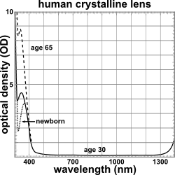
Lens Vertebrate Anatomy Wikipedia

Optics Iii Frame Measurements Flashcards Quizlet

5 1 Physics Of The Eye And The Lens Equation Douglas College Physics 1207

Lens Vertebrate Anatomy Wikipedia

Lens Vertebrate Anatomy Wikipedia

Lens Gene Vision

Schoolphysics Welcome

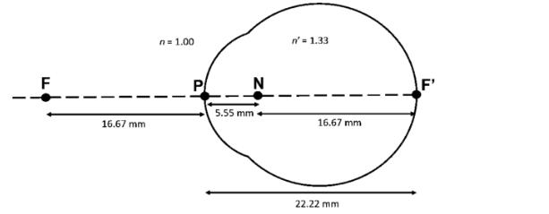
Schematic Eye And Reduced Eye Optography

5 2 Vision Correction Douglas College Physics 1207

Lens Vertebrate Anatomy Wikipedia

Astronomical Optics Part 5 Eyepieces